Echocardiography
Overview
Echocardiography, often called an “echo,” is a test that uses ultrasound waves to create moving pictures of the heart and the large blood vessels around it, like the aorta. It is painless, safe, and gives doctors important information in real time, without using radiation. Echocardiography is a special variant of ultrasound, and much like abdominal ultrasound uses the same ultrasound machine and principles.
How It Works
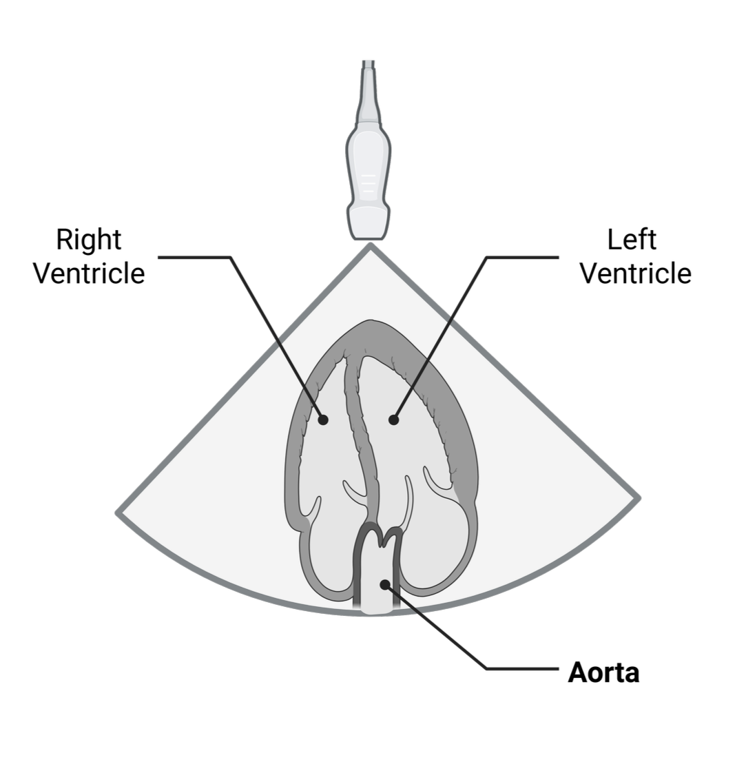
During an echocardiogram, a small device called a transducer (Ultrasound Probe) is placed on your chest or moved gently over the skin. This device sends out sound waves that bounce off the heart and blood vessels, creating images on a screen. There are several types of echocardiograms. The most common is the “transthoracic” echo, where the probe stays on the outside of your chest. Sometimes, a “transesophageal” echo is needed, in which the probe goes into the food pipe (esophagus) to get a closer look at the aorta and heart.

When It’s Used
Doctors use echocardiography to:
Check heart function and heart valves.
Look for fluid around the heart.
Evaluate the size of the aorta, as well as find local damage, such as dissection.
Help find causes of chest pain, shortness of breath, heart failure, fainting, or abnormal heart sounds.
In people with known or suspected aortic disease, an echo might be one of the first tests performed because it is non-invasive and gives quick results.
What It May Not Find
While echocardiography is very helpful, it has some limitations:
It cannot show the whole thoracic aorta, especially the parts in the chest far from the heart.
Small tears, ulcers, or some types of aortic disease may be hard to see if they are not close to the heart.
Extra body tissue, lung problems, or surgical scars may block the sound waves, making the images less clear.
Some changes inside the aorta or its wall layers may require additional scans, such as CT or MRI, for a full assessment.
Complications of Using Echocardiography
Echocardiography is one of the safest heart tests. For the standard test done on the chest, complications are extremely rare. With the transesophageal echo, people might feel throat discomfort, may react to sedation, or, very rarely, have injury to the esophagus. Your medical team will explain the risks before the test.
How It Is Useful in Aortic Disease
Echocardiography is especially valuable for:
Detecting aortic aneurysms and measuring their size.
Identifying aortic dissections close to the heart.
Checking the function of the aortic valve (which may be involved in aortic disease).
Monitoring changes over time after treatment or surgery.
Because echo can show both the heart and key parts of the aorta live and in motion, it often helps doctors quickly make decisions about diagnosis and treatment. In emergencies, such as suspected aortic dissection, an echo can save precious time by providing instant information at the bedside.